


 Sign Out

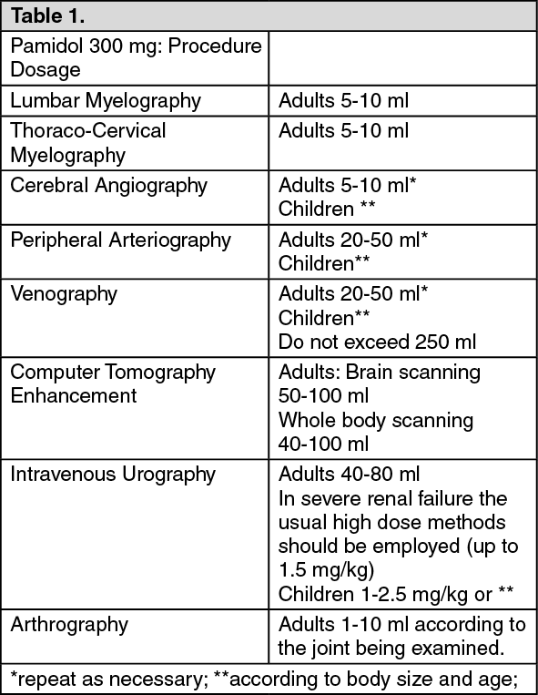 Click on icon to see table/diagram/image
Single injection volume depends on the vascular area to be examined.
Elderly: dosage as for adults. The lowest effective dose should be used.
Method of administration: No other drugs should be mixed with the contrast medium.
Lumbar myelography: A slow sub-arachnoid injection is made through a fine lumbar puncture needle into one of the lower lumbar interspinous spaces (L3-L4 or L4-L5). Optimum contrast appears immediately after injections and films should be obtained promptly.
Thoraco-cervical myelography: Following a slow sub-arachnoid injection the patient should be turned on his side and tilted 10°-20° head down under fluoroscopic control. In this manner it is possible to control movement of the contrast medium column into the dorsal region.
If the cervical region is to be examined, the contrast medium should be run into the cervical region first, before the examination of the dorsal areas where it is progressively diluted.
Iopamidol may also be injected sub-occipitally or by lateral cervical puncture technique. Care should be taken to ensure that the contrast medium does not move intracranially.
Following intrathecal use, the patient should rest with the head and chest elevated for one hour and be kept well hydrated. Thereafter, he/she may ambulate carefully but bending down be avoided. If remaining in bed, the head and chest should be kept elevated for 6 hours. Patients suspected of having a low seizure threshold should be observed during this period.
Cerebral angiography: Any of the current techniques is suitable for radiological visualization of the cerebral vasculature with Iopamidol 300. Carotid and vertebral angiography, performed by catheterization or percutaneous injection techniques, require rapid injection, which, if necessary may be repeated.
Peripheral arteriography and phlebography (venography): Percutaneous injection into the appropriate blood vessel is used for visualization of peripheral arteries and veins.
Computer tomography enhancement: Contrast enhancement for brain scans can be achieved between one and three minutes after i.v. injection. Iopamidol 300 is also used for total body scanning examinations after i.v. administration as a bolus, as a drip infusion or by a combination of the two methods.
Urography: The contrast medium is injected intravenously and rapidly eliminated through the kidneys. In patients with severe renal failure, high dose urography should be used.
Arthrography: Visualisation of joint cavities and articular surfaces can be achieved by either single or double contrast examination.
Mode of Administration: Intravascular Injection.
755.53 mg: (See Table 2.)
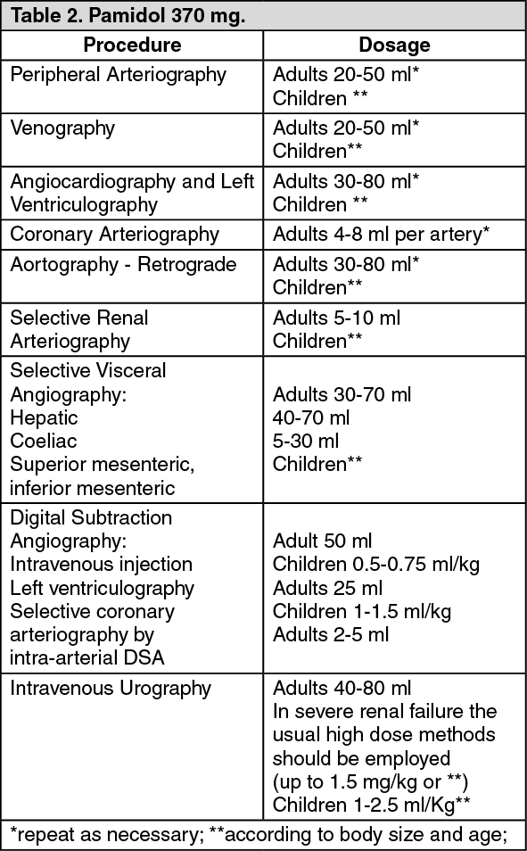 Click on icon to see table/diagram/image
Do not exceed 250 ml. Single injection volume depends on the vascular area to be examined.
Elderly: dosage as for adults. The lowest effective dose should be used.
Method of administration: No other drugs should be mixed with the contrast medium.
Peripheral arteriography and phlebography (venography): Percutaneous injection into the appropriate blood vessel is used for visualisation of peripheral arteries and veins.
Angiocardiography, left ventriculography, selective coronary arteriography: Iopamidol may be administered by rapid injection through a catheter into a suitable peripheral artery or vein. It can also be introduced under pressure through a cardiac catheter into any of the heart chambers, or injected into large vessels for immediate visualisation. The contrast medium may also be administered during selective catheterisation of the coronary arteries.
Aortography: The contrast medium may be introduced directly by intra-arterial injection (retro-grade method) for visualisation of the aorta and its main branches.
Selective visceral angiography: Visualisation can be achieved by selective catheterisation and injection into the hepatic, coeliac or mesenteric arteries.
Digital subtraction angiography: For cardiac imaging the contrast medium may be administered intra-arterially by selective catheterisation to provide subtracted images. Iopamidol 370 injected intravenously either centrally or peripherally is also recommended for use in this modality.
Urography: The contrast medium is injected intravenously and rapidly eliminated through the kidneys. In patients with severe renal failure, high dose urography should be used.
Mode of Administration: Intravascular Injection.